Cell death by sliding
Drs. Wilfred T. Tysoe & Nicholas D. Spencer | TLT Cutting Edge October 2019
Shear stresses above ~100 Pa accelerate the rate of programmed cell death of the epithelial cells coating the surface of the eye.
The epithelium comprises a thin layer of cells that covers and protects many organs in the body, for example the surface of the eye (the cornea). The corneal epithelial cells are coated by a thin liquid film that facilitates nutrient transport, provides protection against microbes, prevents dehydration and helps to lubricate the eye to reduce shear stresses during blinking to 20 to 40 Pa. When the eye is stressed and becomes inflamed, such as in “dry eye” syndrome, the epithelial cells respond by releasing small protein signaling molecules, known as cytokines. These molecules trigger a number of events and recruit immune cells as part of a process called inflammation.
Apoptosis is a cellular process that is often called organized cell death and is used to break apart a cell into small packets of proteins to be cleared from the tissue as a form of maintenance (
see Figure 1); on average we lose between 50 and 70 billion cells each day in this way. The finding that high frictional shear stresses acting on epithelial cells could trigger the release of cytokines (
1) led to the hypothesis that shear stress also may be capable of initiating apoptosis, but whether this indeed takes place, and the stresses at which this might occur, is unknown.
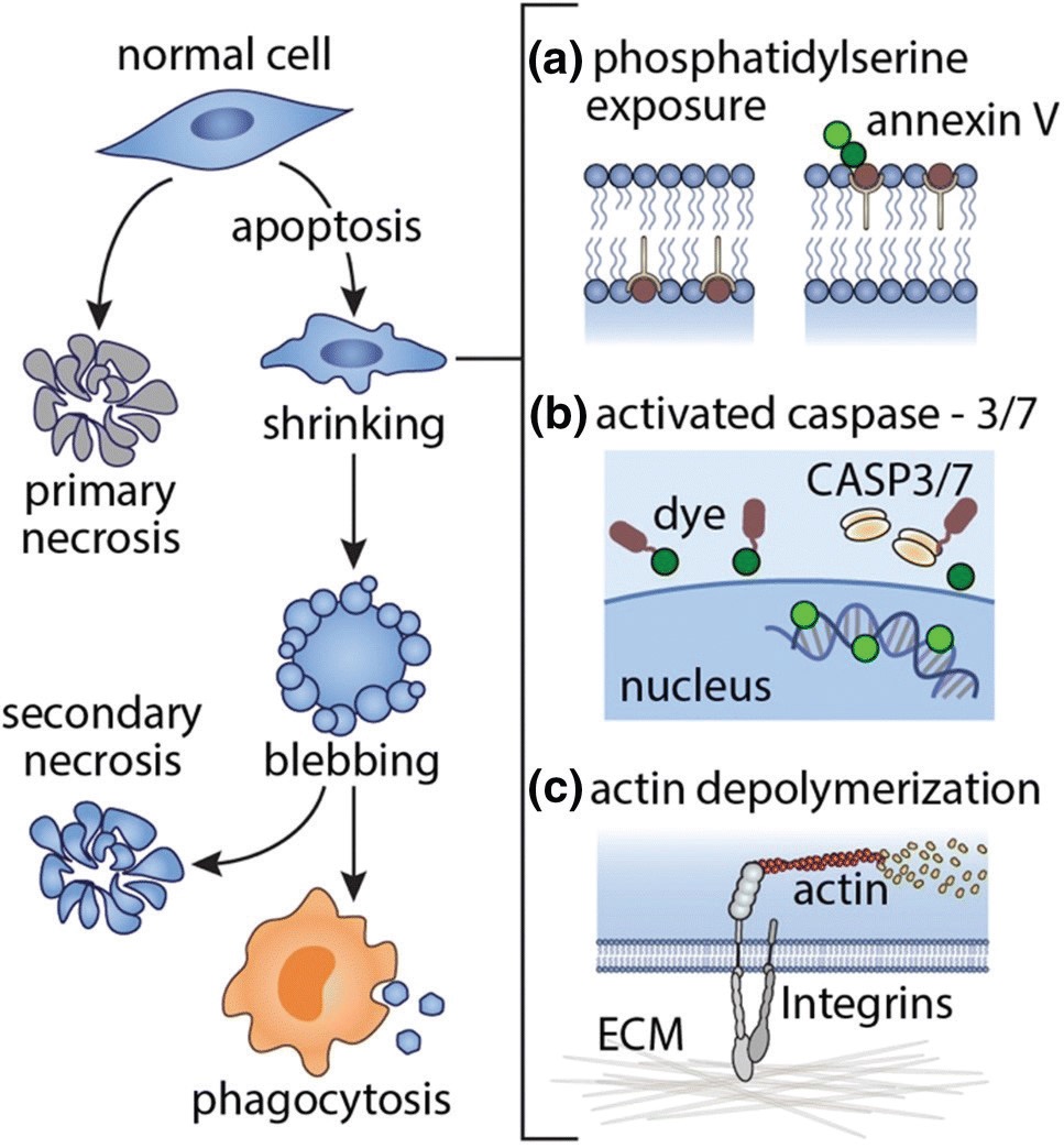 Figure 1. Cell death processes in multicellular organisms include apoptosis and necrosis. Apoptosis is a programmed cell death mechanism that generally involves blebbing (the formation of small blisters) and packaging of the cellular constituents for subsequent phagocytosis (digestion). The hallmarks of apoptosis include (a) phosphatidylserine translocation to the outer cell membrane surface, (b) caspase-3/7 activity, (c) actin depolymerization. (Published with permission from Ref. 2.)
Investigating the influence of shear stress on the extent of apoptosis requires a combination of delicate and precise tribological measurements with the ability to grow and assay carefully prepared biological systems. Professor Angela Pitenis and George Degen from the University of California Santa Barbara collaborated with professors Greg Sawyer and Padraic Levings and post-doctoral researcher Juan Manuel Urueña to monitor the onset of apoptosis for a monolayer of corneal epithelial cells rubbed by a polyacrylamide (pAAm) hydrogel probe. The tribometer measured the normal and friction forces during sliding and was equipped with an epiflourescence microscope to follow the degree of apoptosis using several specific fluorescent probes (
see Figure 1a-c).
Initial experiments showed that shear stresses above ~80 Pa enhanced the extent of apoptosis. To more precisely gauge the shear stresses needed to initiate apoptosis in a single pass, “ramped” loading experiments were carried out in which the load was varied as a function of position on the sample. Here the spatial variation in the extent of apoptosis, measured from fluorescence images (
see Figure 2a), showed that only a few cells had undergone apoptosis for shear stresses below ~80 Pa but then increased linearly for higher shear stresses (
see Figure 2b). Control experiments confirmed that the effect was due to the variation in shear stress, not the effect of higher loads.
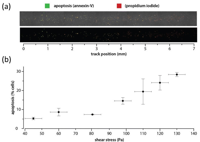 Figure 2. A “ramped” loading experiment was performed to explore whether or not a single pass could initiate apoptosis. (a) Phase-contrast image with overlaid fluorescence (top) and isolated fluorescence channels (bottom). (b) Percent apoptosis versus shear stress with associated uncertainties. Experimental conditions: initial contact radius, a0 ≈ 400 µm, v = 1 mm/s; stroke length, l = 7 mm; number of cycles, n = 1/2 (unidirectional); duration, t ≈ 7 s; average friction coefficient, µ ≈ 0.07. (Published with permission from Ref. 2.)
The authors also pointed out that the critical shear stress required to induce apoptosis is close to the value occurring naturally during blinking or when the eye moves. It seems that nature functions at the limits of the operating conditions without the safety factors that would be included in man-made, engineered systems!
FOR FURTHER READING
1.
Pitenis, A.A., Urueña, J.M., Hart, S.M., O’Bryan, C.S., Marshall, S.L., Levings, P.P., Angelini, T.E., Sawyer, W.G. (2018), “Friction-Induced Inflammation,”
Tribology Letters,
66, 81.
2.
Hart, S.M., Degen, G.D., Urueña, J.M., Levings, P.P., Sawyer, W.G., Pitenis, A.A. (2019), “Friction-Induced Apoptosis,”
Tribology Letters,
67, 82.
Eddy Tysoe is a distinguished professor of physical chemistry at the University of Wisconsin-Milwaukee. You can reach him at wtt@uwm.edu.
Nic Spencer is professor of surface science and technology at the ETH Zurich, Switzerland, and editor-in-chief of STLE-affiliated Tribology Letters journal. You can reach him at nspencer@ethz.ch.