Hip surface science
Drs. Wilfred T. Tysoe & Nicholas D. Spencer | TLT Cutting Edge August 2009
A new paper gives us a better understanding of how articular joints are lubricated.
www.canstockphoto.com
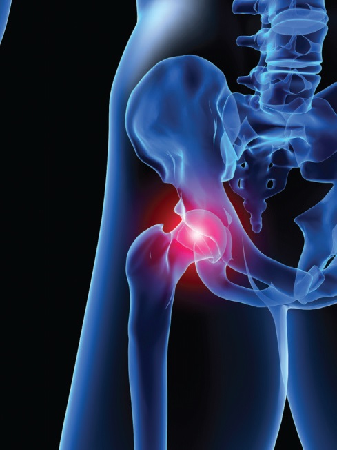
One of the most astonishing tribological systems in nature is that found in our hips and other articular joints. These joints show exceedingly low sliding friction and contain their own lubricant-producing machinery. In most cases they last for many decades without significant wear.
Obviously there are good reasons for trying to understand how these remarkable tribosystems work, not least because our aging population is reaching the end of the useful life of their hip or knee joints in ever-increasing numbers.
Over the years there has been a multitude of different mechanisms invoked to describe the lubrication of articular joints. These have included boundary lubrication, elastohydrodynamic lubrication, microelastohydrodynamic lubrication, squeeze-film lubrication, “weeping” lubrication, “boosted” lubrication, electrostatic lubrication, biphasic lubrication, brush lubrication and gel lubrication to name but a few! In fact, many of these mechanisms may be involved to a greater or lesser extent, or in combination, under different conditions of joint use.
Nevertheless, it is clear that in many situations joints are operating from a standstill. Therefore, some form of boundary lubrication will certainly play a role, and the cartilage surface properties will be highly relevant.
In a recent article published in
Tribology Letters, Rowena Crockett of the Swiss Federal Laboratories for Materials Testing and Research in Dübendorf, Switzerland, focused specifically on boundary lubrication of articular joints (
1). In particular, she handles the delicate issue of the cartilage surface.
This is a trickier topic to investigate than might be expected, since studies of explanted cartilage have been prone to artifacts, and the near-surface region is a highly complex, layered structure. Almost all investigators agree on the components present, however. These include phospholipids (the building blocks of membranes), hyaluronan (a charged, high molecular-weight sugar chain), lubricin (a protein with sugar side-chains, or glycoprotein) and collagen, the main protein of connective tissue.
Working through the many studies that have been published over the last few decades, Crockett put together a view of the cartilage surface that seems consistent with much of the evidence, even though this has previously been interpreted in many different and conflicting ways. Starting inside the bulk cartilage, the cartilage cells, or chondrocytes, are embedded in a sea of collagen and supramolecular structures containing proteins, hyaluronan and glycoproteins, known as aggregate. This bulk structure starts near the bone and goes out to the near-surface region, but we are not at the sliding or articular surface yet. On top of this collagen-rich phase is a hydrophilic layer, or possibly layers, probably consisting of lubricin and very closely related molecules, complexed with hyaluronan. This sugary structure forms the articular surface.
Finally, on top of the articular surface is a dilute, gel-like network of hyaluronan and lipids, which allows facile diffusion of molecules in and out of the cartilage below. Lipids have been shown to dramatically reduce the viscosity of hyaluronan, so this may be the origin of the lubricious, low shear-strength layer present on top of the cartilage surface.
This outer, gel-like surface (or “superficial layer”) of the cartilage is delicate and highly susceptible to drying out and becoming hydrophobic—a phenomenon that led to the hypothesis that cartilage itself is hydrophobic. But as we have seen, the situation is far more complex than this. Methods that have been used to image cartilage surfaces using electron microscopy generally remove the superficial layer, the articular surface, or both, to expose the bulk cartilage. This also has led to inferences based on bulk cartilage morphology, which probably have little to do with the actual sliding surface properties.
Cartilage clearly has a far more complex structure than we are used to in engineering systems, and it works extremely well. Let us hope that a greater understanding of its structure and function will lead to new ways to deal with diseases of the joints and possibly even new directions for the design of materials for low-friction mechanical systems.
REFERENCE
1.
Crockett, R. (2009), “Boundary Lubrication in Natural Articular Joints,”
Tribology Letters,
35 (2), pp. 77-84.
 Eddy Tysoe is a Distinguished Professor of Physical Chemistry at the University of Wisconsin-Milwaukee. You can reach him at wtt@uwm.edu
.
 Nic Spencer is professor of surface science and technology at the ETH Zurich, Switzerland. Both serve as editors-in-chief of STLE-affiliated Tribology Letters journal. You can reach him at nspencer@ethz.ch
.